Figure 11.

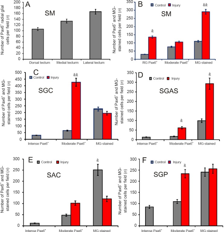
Number of Pax6+ cells, methyl green (MG)-stained cells and radial glial cells in the tectum of intact trout and 1 week after unilateral eye injury (UEI).
(A) Number of Pax6+ radial glial cells in the stratum marginale (SM) of the dorsal, medial, and lateral zones of tectum 1 week after UEI. (B) Number of Pax6+ radial glia cells, moderately Pax6-labeled cells, and Pax6– cells in the SM of tectum. (C) Stratum griseum centrale (SGC) (see designations in B). (D) Stratum griseum et album superficiale (SGAS). (E) Stratum album centrale (SAC). (F) Stratum griseum periventriculare (SGP). (B–F) Data are expressed as the mean ± SD (n = 5 in each group); one-way analysis of variance (ANOVA) followed by the student-newman-keuls post hoc test was used to determine significant difference between control animals and animals subjected to UEI for 1 week. &P < 0.05, &&P < 0.01, vs. control group.
