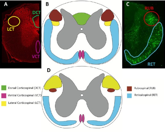
Figure 3.

Comparison of spinal cord tracts.
Transverse sections of the rat (A-C) and human (D) demonstrating the approximate locations and sizes of the corticospinal, rubrospinal, and reticulospinal tracts. Rats were injected with an anterograde tracer, biotinylated dextran amine, into the motor cortex (A), reticular formation and red nucleus (B). Injections performed according to Hellenbrand et al. (2013). The corticospinal tract (CST) has larger lateral fibers in humans (yellow), while there is no dorsal CST in humans as compared to rats, who have a large dorsal CST (green). Both rats and humans have a ventral CST (pink). The rubrospinal tract (red) is prominent in rats, but largely reduced in humans, only passing through the upper cervical levels. All species express the reticulospinal tract (blue) prominently, with slight variations in location and size. High order non-human primates more closely resemble humans; however, tract size and exact location varies between non-human primate species. Adapted from Silva et al. (2013) and Watson et al. (2009).
