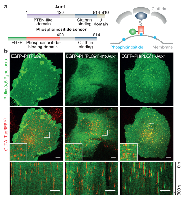
Figure 1 |. Cellular localization of phosphoinositide-specific, auxilinl-based PtdIns(4,5)P2 sensors.
a, Left, domain organization of mammalian Auxl and of fluorescently tagged Auxl-based phosphoinositide sensors. Right, diagram of sensor-coat association. b, Localization of a general PtdIns(4,5)P2 sensor (EGFP-PH(PLCδl)), a mutated Auxl-based PtdIns(4,5)P2 sensor defective in binding PtdIns(4,5)P2 (EGFP-PH(PLCδl)-mt-Auxl), and a wild-type Auxl-based PtdIns(4,5)P2 sensor (EGFP-PH(PLCδl)-Auxl). Top, distribution of PtdIns(4,5)P2 sensor at a single time point; middle, CLTA-TagRFP superposed on PtdIns(4,5)P2 sensor (green), including enlarged region (square box); bottom, corresponding kymographs from 300-s time series imaged every 2 s by spinning-disk confocal microscopy. EGFP channel in the enlarged regions and kymographs shifted laterally by six pixels. Images are representative of at least three independent experiments. Scale bars, 5 μm.

