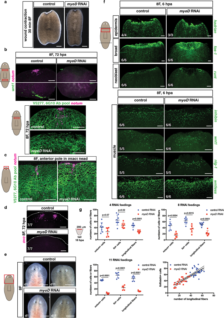
Extended data Figure 4. myoD is required for regeneration.

a, Normal wound contraction in myoD(RNAi) animals, trunk fragments are shown 30 minutes after amputation (15 animals, 3 experiments). b, Lack of anterior (notum+) and posterior (wnt1+) pole cells (top) and BWM structure (bottom) at 72hpa during regeneration in myoD(RNAi) animals, or c, in an uninjured animals (10 animals/group, 3 experiments). Scale bars, 50 μm. d, Neoblasts did not specify into eye progenitors (ovo+) in myoD(RNAi) animals at 72hpa (1 experiment). e, Homeostatic eye replacement at 11 days following eye resection in myoD(RNAi) animals (10 animals/group, 1 experiment). Scale bars, 500 μm. f, Normal epidermis, neoblast and muscle expression of wound-induced genes in myoD(RNAi) animals 6hpa. g, Graphs show reduced numbers of notum+ and follistatin+ cells in myoD(RNAi) animals at 18hpa, and longitudinal fibers after different numbers of RNAi feedings. Cartoon shows the region counted. Linear correlation between follistatin+ cells and longitudinal fibers. Regression coeficient, R2= 0.6928. Two-tailed Student-t test was performed. p-values are shown in graphs. Mean ±SD are shown in all graphs. Bottom left number: animals with phenotype out of total tested. Anterior, up. Scale bars, 100 μm unless indicated.
