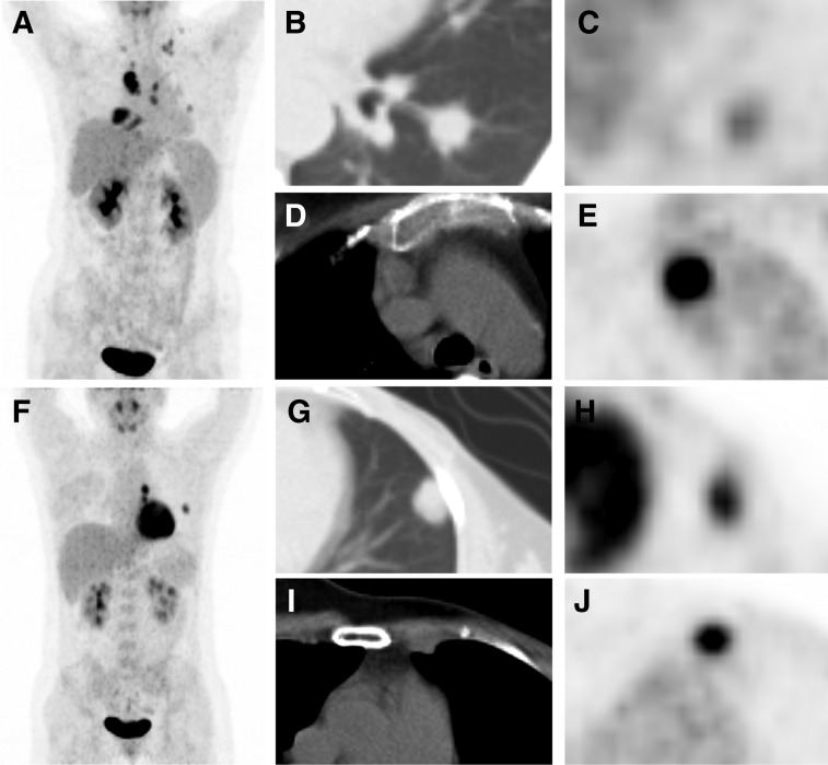
Figure 5.
Representative images. (A–E): A 45‐year‐old female patient with metastatic triple‐negative breast cancer (mTNBC) underwent 18F‐fluorodeoxyglucose (18F‐FDG) positron emission tomography (PET)/computed tomography (CT) scan (A, maximum intensity projection [MIP] image). We detected that the left lung lesion had the lowest 18F‐FDG uptake in all metastatic lesions (B, CT image; C, PET image, minimum FDG uptake across all lesions [MIN]: maximum standard uptake value [SUVmax] = 2.0), whereas the mediastinal lymph node lesion had the highest uptake (D, CT image; E, PET image, maximum FDG uptake across all lesions [MAX]: SUVmax = 15.3). Therefore, heterogeneity index (HI)‐total lesions (‐T) of this patient was 7.7, and she had a progression‐free survival (PFS) of 3.3 months and an overall survival (OS) of 7.4 months. (F–J): A 35‐year‐old female patient with mTNBC underwent 18F‐FDG PET/CT scan (F, MIP image). We detected that the left lung lesion had the lowest 18F‐FDG uptake in all metastatic lesions (G, CT image; H, PET image, MIN: SUVmax = 5.9), whereas the left internal mammary lymph node lesion had the highest uptake (I, CT image; J, PET, MAX: SUVmax = 8.8); Therefore, HI‐T of this patient was 1.5, and she had a PFS of 10.6 months and an OS of 15.0 months.

