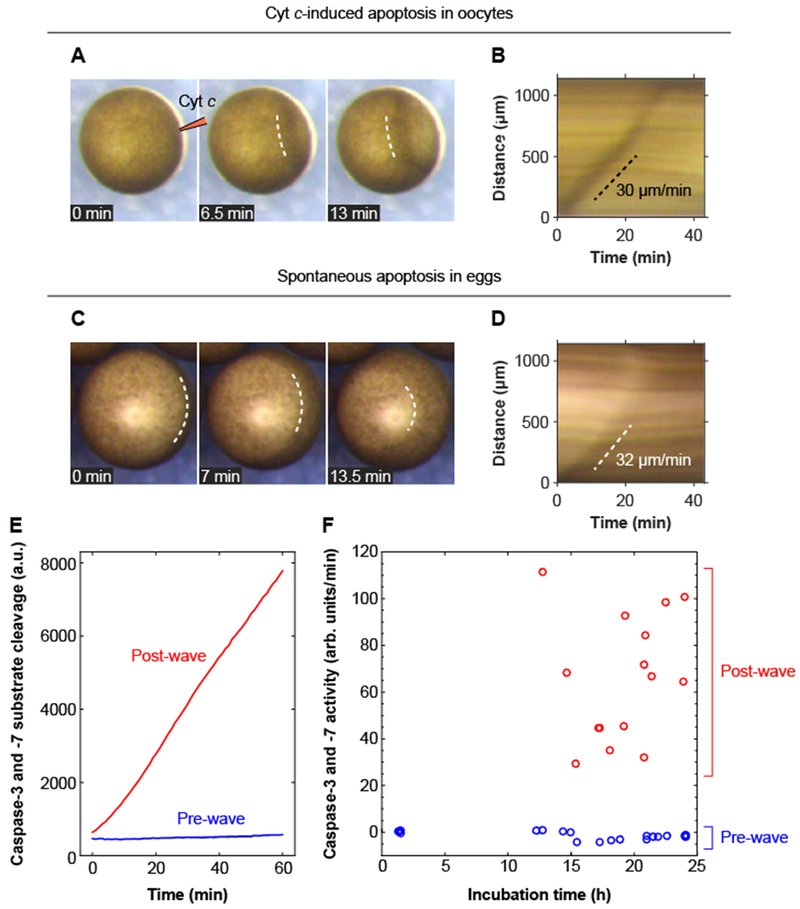
Fig. 4. Apoptotic trigger waves in intact oocytes and eggs.

(A, B) Injection of immature Stage VI oocytes with cytochrome c (10 nl of 1 mg/ml cytochrome c) causes a wave of pigmentation changes to spread from the injection site to the opposite side of the oocyte. Panel A shows one example of this surface wave in montage form; panel B shows the kymograph. Two other examples of these waves are shown in movie S9. (C, D) Surface waves occur in spontaneously dying eggs. Panel C shows an example of this wave in montage form, and panel D shows the kymograph. (E) Caspase-3 and/or caspase-7 assays for one pre-wave and one post-wave egg. (F) Caspase-3 and/or −7 activities for eggs pre- and post-wave. The data are from 19 pre-wave eggs and 15 post-wave eggs.
