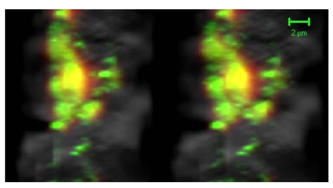
Figure 4.
With the confocal mode, GH protein was observed as a 655 nm emission signal using Qdot 655 (red), and GH mRNA was observed as a 605 nm emission signal using Qdot 605 (green). When GH mRNA and protein were located in the same or adjacent places, their signals were detected in the mixed color images (yellow) (stereo-images, bar = 2 μm [33]).

