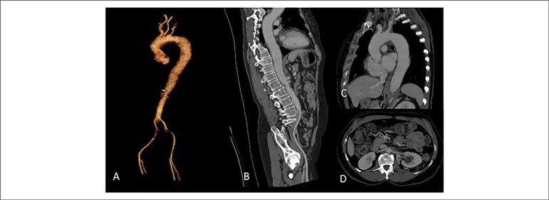
Figure 2.
A) Thoracic and abdominal aorta in 3D reconstruction. B) Aorta seen in the sagittal view, showing diffuse thickening of the entire wall with a narrowing area in the infra-renal aorta. C) Thoracic aorta with parietal thickening and luminal reduction in the origin of left subclavian artery. D) Thickening at the origin of the renal arteries, without significant obstruction characterization.

