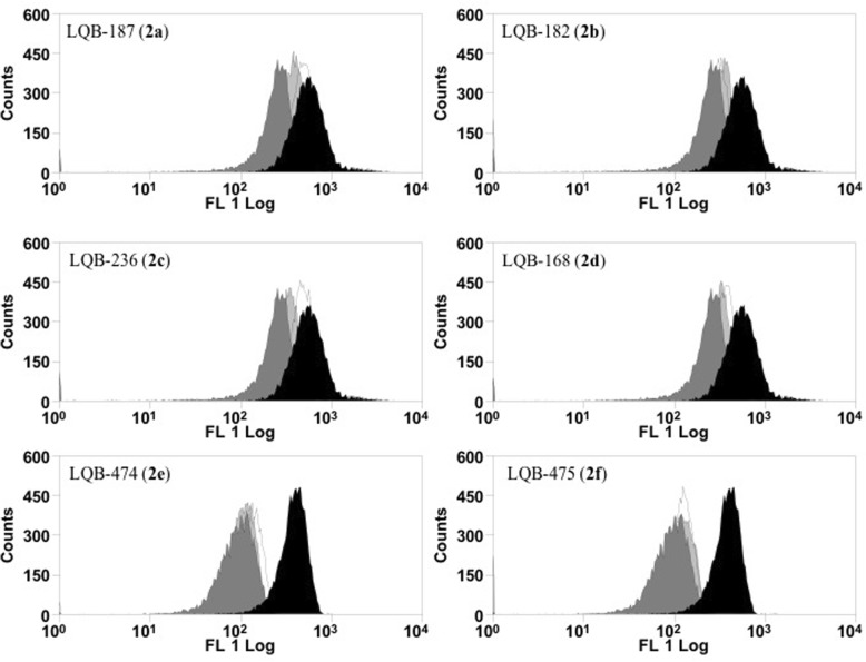
Fig. 3.
L. infantum promastigotes altered mitochondrial membrane potential after treatment with pterocarpanquinones. Promastigotes of L. infantum (5 × 10⁶ cells/mL) were cultured in the presence of 0–2.5 μM of derivatives at 26 °C. After 4 h, the parasites were incubated for 15 min with 10 μg/mL rhodamine 123 (Rh123). Data acquisition and analysis were performed using a FACSCalibur flow cytometer. The FCCP 20 μM was used as positive control. Black (control); White (1.25 μM); Light Gray (2.5 μM) and Dark Gray (FCCP)

