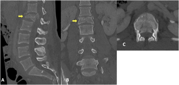
Fig. 3.

A-C (A) Sagittal, (B) coronal, and (C) axial view CT scans show an acute L1 burst fracture (arrows in Illustrations A and B) with retropulsion. There is loss of vertebral body height seen on the sagittal and coronal views with fragment retropulsion evident on the axial view.
