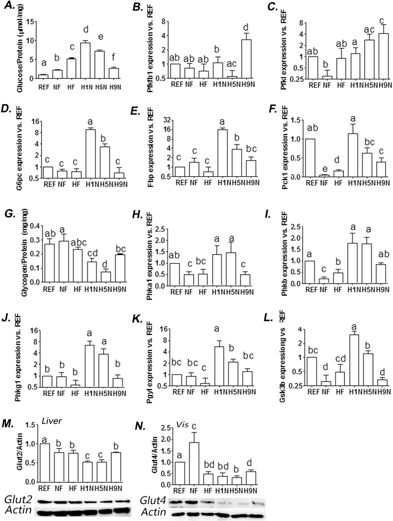
Figure 4. The H9N, but not the H5N and H1N diets, normalized offspring glucose homeostasis broken by maternal HF diet.

4A. Amount of hepatic glucose content after 12-week exposure of postweaning HF diet. Hepatic glucose level was measured using an Amplex red glucose assay kit (Thermo Fisher Scientific Inc, #A22189). 4B-F. Hepatic expression of genes involved in glycogenesis and gluconeogenesis, and glycolysis was measured by real-time PCR. 4G. Amount of hepatic glycogen content after 12-week exposure of postweaning HF diet. Hepatic glycogen level was measured using a glycogen assay kit (Thermo Fisher Scientific Inc, #MAK016-1KT). 4H-L. Hepatic expression of genes involved in inhibiting glycogenesis was measured by realtime PCR. 4M. Hepatic Glut2 expression was detected by Western blot analysis. Relative amounts of Glut2 were expressed as the ratio Glut2/Actin. 4N. Adipocyte Glut4 expression was detected by Western blot analysis. Relative amounts of Glut4 were expressed as the ratio Glut4/Actin. The results are presented as Mean ± SE, n= 3-7. Significance (p < 0.05) is presented by different characters comparing among groups in each tissue.
