Figure 4.

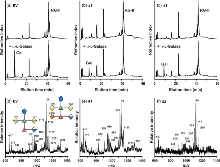
α‐l‐galactosidase treatment confirms that the rhamnogalacturonan‐II (RG‐II) from the hairpin (hp) GGLT1 lines is deficient in α‐l‐galactose. (a)–(c). The high‐performance anion exchange chromatography with pulsed amperometric detection profiles of untreated (top) and α‐l‐galactosidase‐treated (bottom) RG‐II from the empty vector (EV) control and hpGGLT1 lines. The elution position of galactose, which was the only monosaccharide detected, is shown. (d)–(f) The negative ion matrix‐assisted laser desorption–ionization time‐of‐flight mass spectrum of side‐chain A generated by selective acid hydrolysis of L‐galactosidase‐treated RG‐II. The predominant oligosaccharide (m/z 1115, 1129, 1143 and 1157) corresponds to side‐chain A lacking L‐galactose. The differences in mass of 14 Da correspond to differences in the extent of O‐methylation of side‐chain A. Only low‐intensity signals (m/z 1277, 1291 and 1305) corresponding to the L‐galactose‐containing side‐chain A were detected. The oligosaccharide structures are represented using a modified Symbol Nomenclature for Glycans nomenclature. See Figure 1 for details.
