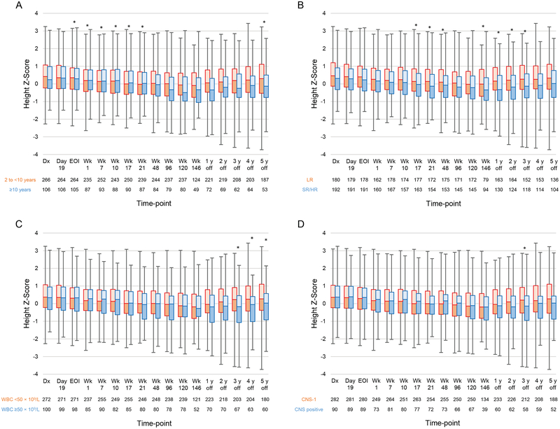
Figure 5.
Longitudinal changes in the median height Z-scores according to (A) age (2 to <10 years vs. ≥10 years); (B) treatment risk (low risk [LR] vs. standard/high risk [SR/HR]; (C) white blood cell count (WBC) at diagnosis (<50×109/L vs. ≥50×109/L); and (D) central nervous system (CNS) disease status at diagnosis (CNS-1 vs. CNS positive). Boxes show scores from the first quartile to the third quartile. The border between the 2 boxes shows the median Z-score. Error bars show minimum and maximum values. Time-points at which there were significant differences between the 2 groups in the mixed model are indicated by asterisks. Abbreviations: Dx, diagnosis; EOI, end of induction; Wk, week.

