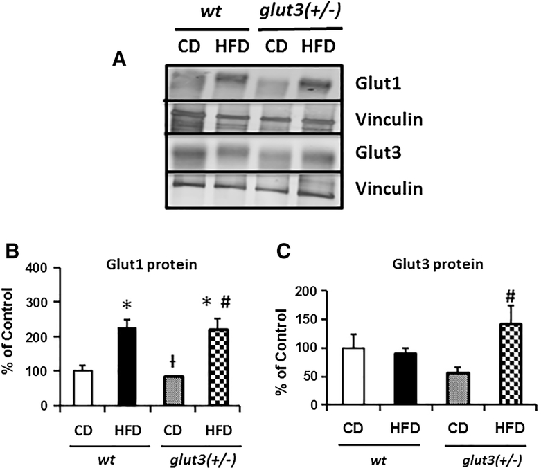
Figure 6. Placental Glucose transporters.
A. Representative Western blots are seen in the inset depicting Glut1 and Glut3 protein bands with vinculin serving as the internal loading control. Below is the graph demonstrating quantification of Glut1 and Glut3 protein (N=8/group/genotype) depicted as a ratio to the vinculin protein and expressed as a percent of the wt ad libitum fed control values. Placental Glut3 protein quantification analysis is as follows: F value (2.9), glut3+/−HFD vs #glut3+/−CD (p=0.006). Placental Glut1 protein analysis is as follows: F value (9.66), wtHFD (p=0.006). glut3+/HFD (p=0.008) vs wtCD, glut3+/−HFD vs glut3+/−CD (p=0.004) and glut3+/−CD vs †wtHFD (p=0.004).

