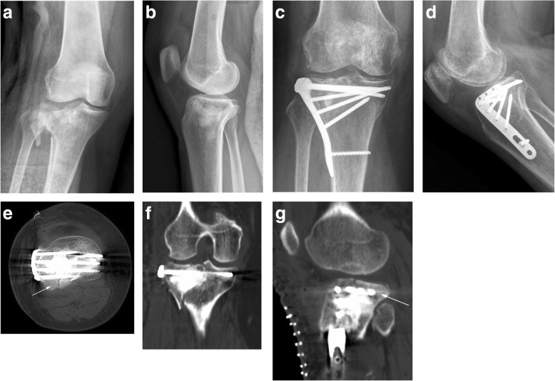
Fig. 2.
A case of type II tibial plateau fracture, female, 60 years old. a, b Preoperative X-ray showed lateral plateau fracture, increased width of the plateau, and posterolateral fracture. c, d Postoperative X-ray showed that the fracture was anatomically reduced and fixed by a lateral locking plate with rafting screws. e–g CT scan showed an anatomical reduction, and the arrow indicated the posterolateral fracture got satisfactory fixation by screws

