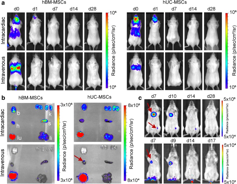
Fig. 6.
Long-term monitoring of human MSCs in BALB/c mice. a Representative BLI of mice administered with 5 × 10⁵ hBM-MSC or hUC-MSC via the IC or IV route. The signal was progressively lost shortly after administration, with no evidence of malignant growth. b Ex vivo bioluminescence imaging of organs within 5 h of administration of the cells. Organs are indicated as the kidneys (k), spleen (s), liver (li), lungs (lu), heart (h) or brain (b). In some occasions, signal foci were seen in the heart of mice that received hUC-MSC IV (red arrow). c BLI images from mice that displayed hUC-MSC signal that persisted beyond day 7 after IV administration (ventral orientation, lower scale). In all cases, the signals had disappeared by day 21 and had not returned by the end of the experiment

