
Figure 3.
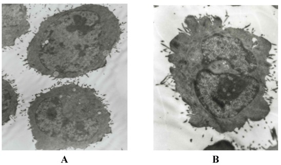
Morphological observation of H22 cells by EM after treatment. The cells were examined under a transmission electronmicroscope (×5000 power, bar = 1 μm). A: normal hepatoma H22 cells; B: karyopyknosis, membrane integrity and formation of apoptotic bodies in high dosage CE-SB group.