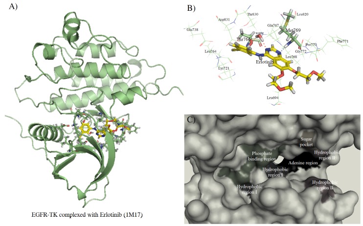
Figure 4.
The complexes of EGFR-TK and erlotinib. (A) Overall structure of TK complexed with erlotinib. (B) The erlotinib and binding residues of kinase domain. (C) The molecular surface representation of the ATP-binding region which consists of adenine region, hydrophobic region I and II, sugar pocket and phosphate binding region.

