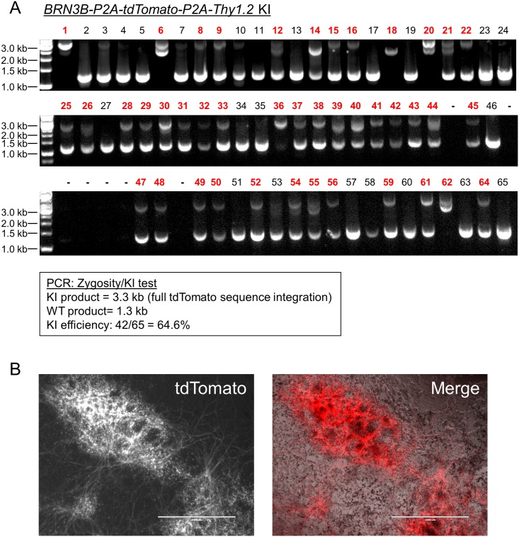
Fig 4. Generation of an RGC reporter line in EP1 hiPSC background using transient puromycin treatment.
(A) PCR zygosity test for KI at the targeted BRN3B locus. Primers spanning the integration region were used to amplify genomic DNA from randomly picked colonies derived from plating single cells. Homozygous insertion of the KI cassette is indicated by a single band at 3.3 kilobase pairs (kb). KI negative clones generate a band of 1.3 kb. Clones producing both bands were scored as heterozygous KI. WT = wildtype. For some of the clones (e.g. lane 6), the KI product is split into two parts due to an incorporation of only one monomer of the tdTomato sequence. (B) Fluorescence and phase microscopy of a differentiated EP1 hiPSC RGC reporter line generated using transient puromycin selection. Cells were imaged on day 29 of differentiation. Scale bar = 1000 μm.

