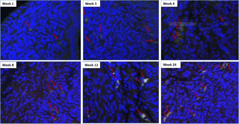
Fig. 1.
OCT4/GFP and CD44 co-staining of mice myometrium. Uterine ages 1, 3, 4, 8, 12, and 24 weeks (40×) are shown. Because Oct-4 was tagged with GFP, the cells expressing Oct-4 emitted green fluorescence. The conjugated CD44 antibody expressed Texas Red Fluorescence. The combination of both Oct-4 and CD44 staining (red and green) is yellow. Here, we show the yellow staining that indicates co-localization of Oct4/GFP and CD44

