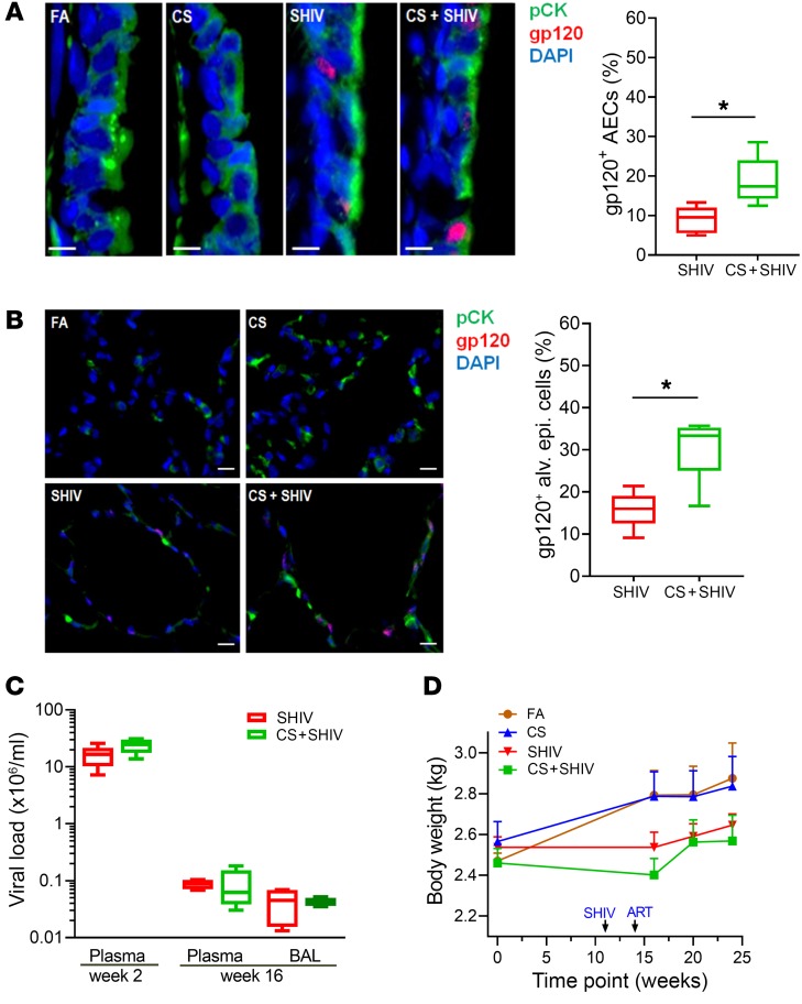
Figure 1. CS increases the number of gp120-immunopositive lung epithelial cells in SHIV-infected and cART-treated cynomolgus macaques.
Formalin-fixed and paraffin-embedded (FFPE) lung tissue sections (5 μm) from each group were immunostained for the epithelial cell marker pan-cytokeratin (pCK) and for HIV-gp120. (A) Representative micrographs of bronchial airway epithelial cells (AECs) showing pCK (green) and HIV-gp120 (red) immunopositivity along with the DAPI-stained nuclei (blue) from each group. Scale bars: 5 μm. Percentages of gp120+ AECs were quantified. (B) Micrographs of alveolar region showing pCK (green) and HIV-gp120 (red) immunopositivity from each group. The percentage of gp120+ alveolar epithelial cells were quantified. Scale bars: 10 μm. Data for A and B are mean ± SEM, n = 7/group; *P ≤ 0.05 by Student’s t test. (C) Viral titers in the plasma and in the plasma and bronchoalveolar lavage (BAL) of SHIV and CS+SHIV-exposed animals at weeks 2 and 16 postinfection, respectively. (D) Body weights of macaques at the baseline and at indicated times during the experiment. Data are mean ± SEM, n = 4–7/group.

