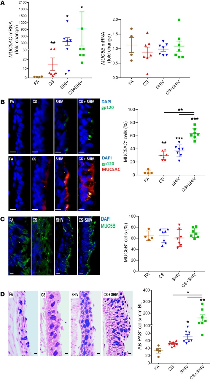
Figure 2. CS exposure and SHIV infection synergistically induce mucous cell hyperplasia and MUC5AC mucin expression.
(A) Quantification of secretory mucin MUC5AC and MUC5B mRNA levels in each group. RNA was isolated from frozen lung tissues and assessed using real-time qRT-PCR. (B) Micrographs of small airways showing MUC5AC (red) and HIV-gp120 (green) immunopositivity from each group, and the percentage of MUC5AC+ epithelial cells. Scale bars: 5 μm. (C) Micrographs of small airways showing MUC5B (green) immunopositivity from each group, and the percentage of MUC5B+ epithelial cells. Scale bars: 5 μm. (D) Representative micrographs of small airways showing mucopolysaccharides stained with AB-PAS. Scale bars: 5 μm. AB-PAS+ cells were quantified per unit length (mm) of basal lamina (BL). Data are mean ± SEM, n = 4–7/group, 1-way ANOVA; *P ≤ 0.05; **P < 0.01; ***P < 0.001.

