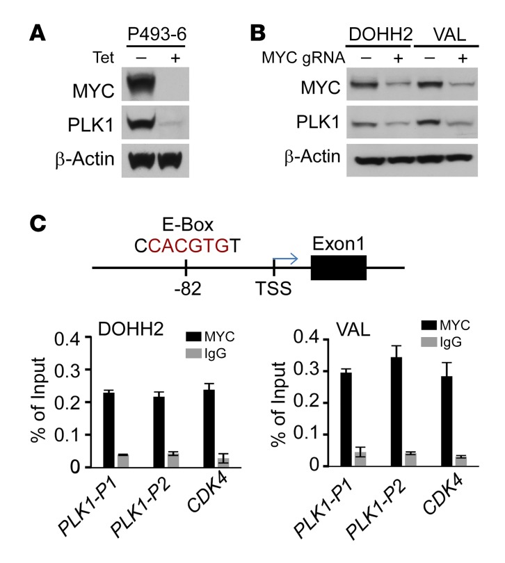
Figure 4. MYC activates PLK1 transcription in DHL.
(A) PLK1 protein levels are dependent on MYC. P493-6 cells were cultured with Tet for 72 hours, and a portion were then deprived of Tet for 24 hours. Levels of MYC, PLK1, and β-actin were determined by Western blot. (B) MYC KD by CRISPR/cas9 gene editing provokes reductions in PLK1 protein in DHL DOHH2 and VAL cells. (C) Upper panel: an E-box site that conforms to the preferred binding site for MYC (CACGTG, PLK1-P1) is located approximately 80 base pairs upstream of the PLK1 transcription start site (TSS). Lower panel: ChIP assays revealed MYC binds to this region of the PLK1 promoter in DHL cells. Binding of MYC to the CDK4 promoter was assessed as a positive control. A and B are representative of 3 independent experiments. Data presented in C show the mean ± SD of at least 3 independent experiments. See complete unedited blots in the supplemental material.

