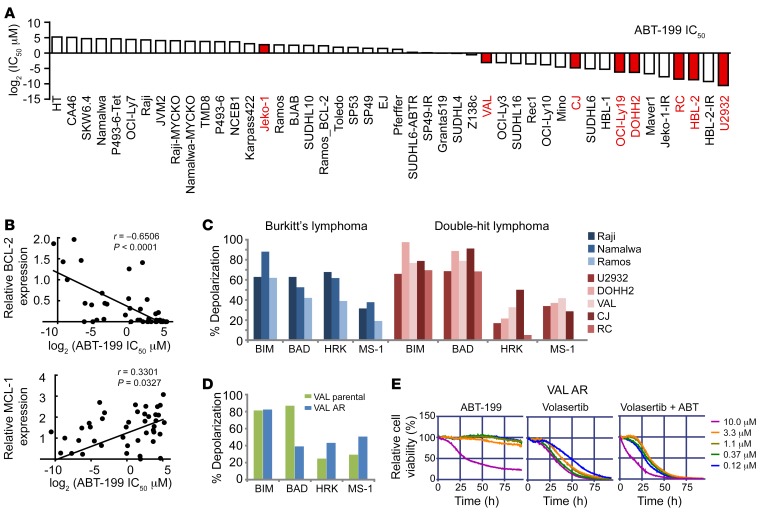
Figure 6. BH3 profiling and sensitivity of DHL and ABT-199–resistant DHL to PLK1 inhibition.
(A) IC50 of the indicated B lymphoma cell lines to ABT-199. DHL/DEL cell lines are highlighted in red. (B) Correlation of BCL-2 and MCL-1 protein levels and ABT-199 IC50. IC50 values were determined using MTT assays, and protein levels were determined by Western blot. (C) BH3 profiling of DHL (red, MYChi/BCL-2hi) and BL (blue, MYChi/BCL-2lo) cell lines showing the dependency of most lines to BCL-2 priming. Increased sensitivity (mitochondrial membrane depolarization) to (BAD-HRK) peptides is indicative of BCL-2 dependency; thus, DHL cells are sensitive to ABT-199. (D) BH3 profiling of ABT-199–resistant DHL cells (VAL_AR) reveals a shift to dependency on MCL-1. (E) Viability of VAL_AR cells treated with ABT-199 (left), volasertib (middle), or both volasertib and ABT-199 (right).

