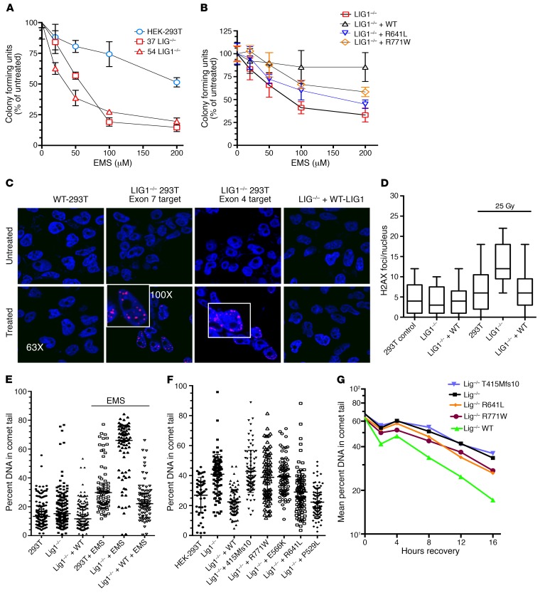
Figure 3. Defective repair of LIG1–/– HEK-293T and mutant cells.
(A) LIG1–/– clones 37 and 54 were more sensitive to EMS, as compared with HEK-293T cells but similar to each other (2 experiments, 6 replicates; mean ± SD; P = 0.03, P = 0.04). (B) EMS sensitivity of clone 37 cells was rescued by complementation with WT LIG1, but only partially with R641L or R771W (3 experiments, 6 replicates; mean ± SD). (C) Responses of HEK-293T cells to γ radiation: WT HEK-293T cells, clone 37 LIG1–/–, and LIG1–/– + WT LIG1 were irradiated (25 Gy), incubated for 3 hours, and inspected for number of γH2AX foci per nuclei (3 experiments). (D) γH2AX foci/nucleus for HEK-293T cells, LIG1–/–, and LIG1–/– cells complemented with WT LIG1, before and after irradiation with 25 Gy. LIG1–/– cells demonstrated increased foci compared with HEK-293T cells (P = 0.0001), with substantial rescue with WT protein: LIG1–/– versus LIG1 –/– + WT, now P = 0.05 (3 experiments; 50 nuclei/condition counted per experiment; mean ± SD). (E) HEK-293T cells, LIG1–/– cells, and LIG–/– cells transfected with WT LIG1, incubated with media alone or 0.5 mM EMS, and examined by comet assay. Data are mean ± SD and percentage of cellular DNA in the comet tail for 79 to 86 cells/condition; performed 3 times. (F) Comparing DNA damage in 0.5 mM EMS-treated HEK-293T cells to LIG1–/–, T415Mfs*10, R771W, E566K cells, P < 0.0001, to R641L cells, P = 0.69. Adding the P529L variant was similar to adding WT LIG1 (differences, P = 0.9). (G) After 16 hours EMS, LIG1–/–, and mutant cells were washed, replated in fresh media, and tested for the differences in DNA damage at intervals. Mean percent shown for each. At 8 hours, differences emerged: HEK-293T versus LIG1–/–, P = 0.007; LIG1–/– versus T415Mfs*10, P = not significant; LIG1–/– versus R641L, P = 0.02; LIG1–/– versus R771W, P = 0.04 (50–88 colonies/condition).

