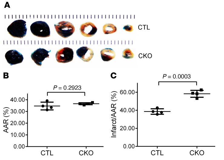
Figure 6. Cardiac Ubqln1 ablation increases infarct size induced by acute myocardial IRI in mice.
Myocardial IRI was produced as described in Figure 5. At 24 hours of reperfusion, the animal was sacrificed, the original coronary ligature was retied, and the heart was subject to retrograde perfusion with 5% phthalocyanine blue to demarcate the original ischemic area or AAR before undergoing TTC staining to reveal the infarct area. (A) Representative pair of TTC staining images. (B) AAR presented is the percentage of LV that was subject to ischemia during the initial LAD ligation phase. (C) Changes in infarct size. Two-tailed Student’s t test. Each dot represents an individual animal.

