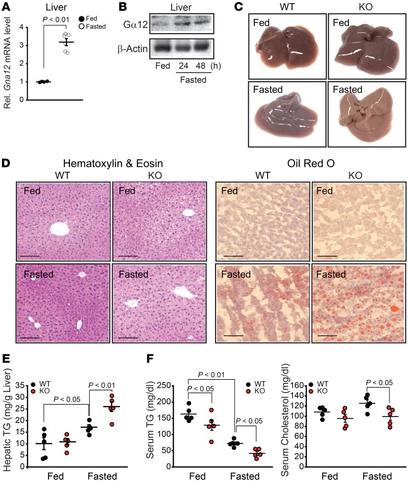
Figure 1. Association of Gα12 signaling with fasting-induced liver steatosis.
(A) qRT-PCR assays for Gna12 in the liver from 10-week-old mice fed ad libitum or fasted for 24 hours (n = 4–6/group). Rel., relative. (B) Immunoblotting for Gα12 in liver homogenates from WT mice fed ND ad libitum or fasted for indicated times. Blots were run in parallel using the same samples. (C) Representative gross appearance of liver tissues from the mice shown in A (n = 3/group). (D) Representative H&E staining (left; n = 5/group) and oil red O staining (right; n = 3/group) of the liver sections. Scale bars: 100 μm. (E) Hepatic TG contents (n = 5/group). (F) Serum TG and total cholesterol levels (n = 5/group). Values represent mean ± SEM. Data were analyzed by 2-tailed Student’s t test (A) or ANOVA, followed by LSD post hoc tests (E and F).

