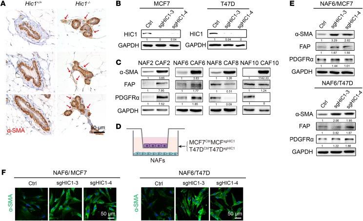
Figure 2. HIC1-deleted BrCa cells induce the activation of stromal fibroblasts in mammary gland.
(A) Representative 3 immunohistochemistry images for α-SMA in mammary glands of 6-month-old mice. Positive staining in the mammary gland stroma of Hic1–/– mice is indicated by red arrows (n = 3 for each group). (B) CRISPR-Cas9–mediated HIC1 deletion in MCF7 and T47D luminal BrCa cells. Cell lysates were analyzed by Western blot with antibodies against HIC1 and GAPDH. sg3 and sg4 represent 2 different interference sgRNA sequences. Ctrl, control. (C) Representative primary NAFs and CAFs isolated from human BrCa tissues. Western blot analysis of cell lysates was performed using antibodies against α-SMA, FAP, PDGFRα, and GAPDH. (D) Schematic showing primary NAFs cocultured with MCF7CtrlMCF7sgHIC1 or T47DCtrlT47DsgHIC1 luminal BrCa cells in a Transwell apparatus (0.4 μm pore size) for 4 days. (E) Western blot analysis of lysates of NAF6 cells that were cocultured with MCF7CtrlMCF7sgHIC1 or T47DCtrlT47DsgHIC1 luminal BrCa cells for 4 days. Antibodies against α-SMA, FAP, PDGFRα, and GAPDH were used. (F) Immunofluorescence staining for the detection of α-SMA expression in NAF6 cells that were cocultured with MCF7CtrlMCF7sgHIC1 or T47DCtrlT47DsgHIC1 luminal BrCa cells for 4 days.

