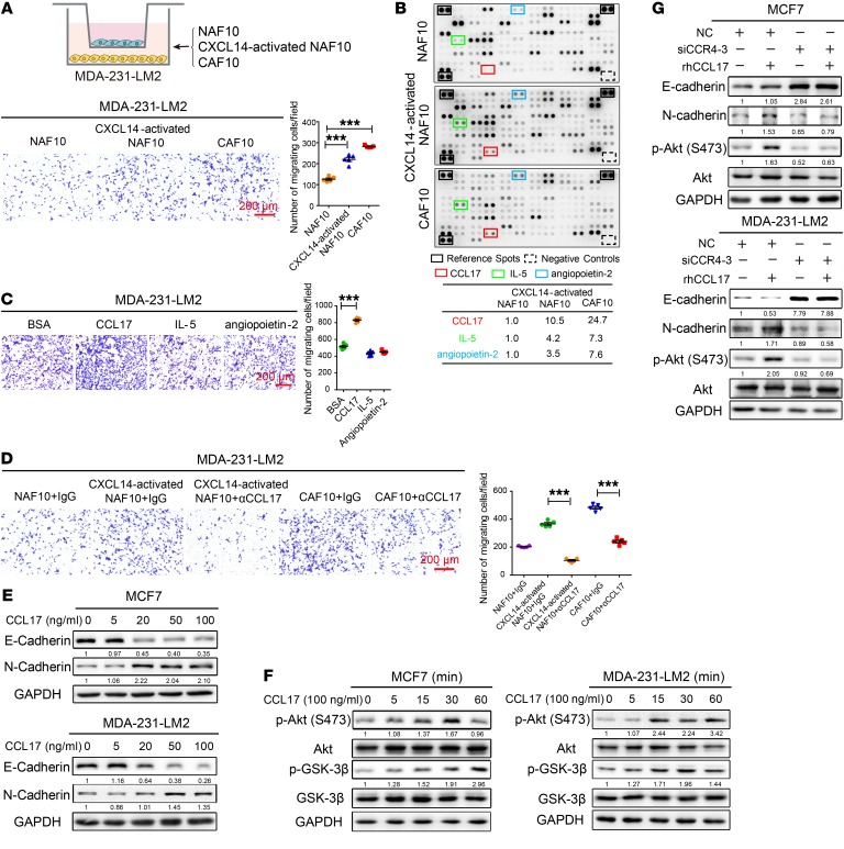
Figure 6. CXCL14-activated fibroblasts induce migration of BrCa cells via CCL17.
(A) Upper panel: schematic showing the coculture of MDA-231-LM2 BrCa cells with primary NAF10, CXCL14-activated NAF10, or primary CAF10 cells in a Transwell apparatus (0.4 μm pore size) for 4 days. Lower panel: Boyden chamber assay of MDA-231-LM2 cells that were treated as above. (B) Upper panel: Human XL Cytokine Array Kits (R&D Systems) were used to measure the levels of 102 cytokines in the CM from diverse fibroblasts. Cytokines that were upregulated in the CM of CXCL14-activated NAF10 and CAF10 cells are indicated by colored boxes; they include CCL17 (red), IL-5 (green), and angiopoietin-2 (blue). Black frames indicate the positive controls, and the dashed boxes indicate the negative controls in each membrane. Lower panel: table showing the relative signal intensities of the 3 selected cytokines noted above. The signal intensities were quantified by densitometry using ImageJ software and normalized to the intensity of the internal positive controls. (C) Boyden chamber assay of MDA-231-LM2 cells plated with rhCCL17, rhIL-5, and rh angiopoietin-2 in the lower chambers at 100 ng/ml for 20 hours. (D) Boyden chamber assay of MDA-231-LM2 cells that were cocultured with primary NAF10, CXCL14-activated NAF10, or primary CAF10 cells in a Transwell apparatus for 4 days in the presence or absence of α-CCL17 (1 μg/ml) or an isotype-matched IgG control. (E) MCF7 or MDA-231-LM2 cells were treated with various concentrations (0–100 ng/ml) of rhCCL17 for 4 days, and lysates of the cells were analyzed by Western blot using antibodies against E-cadherin, N-cadherin, and GAPDH. (F) MCF7 or MDA-231-LM2 cells were treated with rhCCL17 at 100 ng/ml for the indicated times (0, 5, 15, 30, and 60 minutes). Cell lysates were analyzed by Western blot with antibodies against p-Akt (Ser 473), Akt, p-GSK-3β (Ser9), GSK-3β, and GAPDH. (G) Knockdown of CCR4 expression by siRNA-3 in MCF7 and MDA-231-LM2 cells in the presence or absence of rhCCL17 at 100 ng/ml for 4 days. Cell lysates were analyzed by Western blot with antibodies against E-cadherin, N-cadherin, p-Akt (Ser 473), Akt, and GAPDH. Data are shown as mean ± SEM. n = 3 independent experiments. ***P < 0.001, 1-way ANOVA followed by Bonferroni’s post hoc test.

