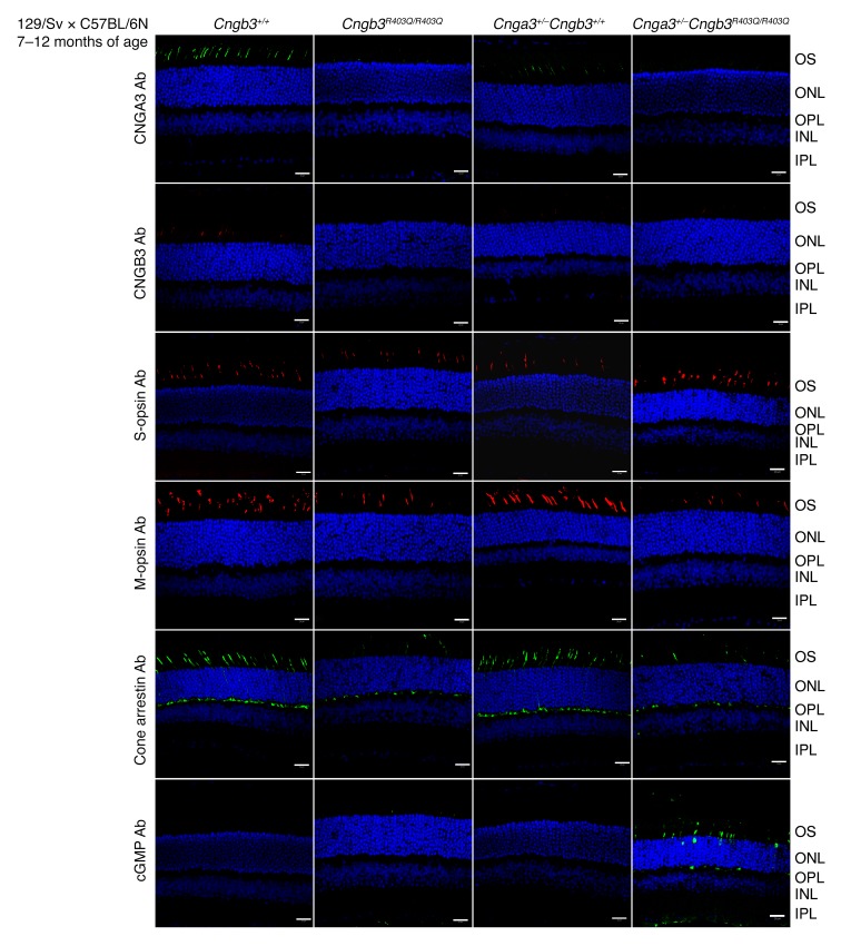
Figure 3. Cone degeneration and reduced CNGA3 and CNGB3 immuno staining in aged Cngb3R403Q/R403Q and Cnga3+/– Cngb3R403Q/R403Q mutants.
Immunofluorescence of cryosections from murine retinas (n = 3 per genotype) at 7–12 months of age (WT, Cnga3+/– Cngb3+/+, Cngb3R403Q/R403Q, and Cnga3+/– Cngb3R403Q/R403Q). Scale bars: 20 μm. S-opsin and cGMP staining with the respective antibodies was performed simultaneously on the same slices and imaged by multicolor laser scanning confocal micrography. Antibodies were diluted as follows: cGMP: 1:3,000; cone arrestin: 1:500; S-opsin: 1:300; M-opsin: 1:300; and CNGB3-antiserum: 1:5,000. INL, inner nuclear layer; IPL, inner plexiform layer; ONL, outer nuclear layer; OPL, outer plexiform layer; OS, outer segment.

