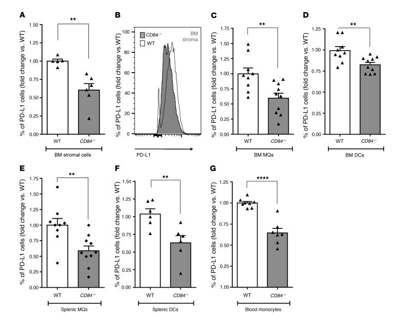
Figure 4. Decreased PD-L1 expression on cells in the microenvironment in the absence of CD84.
(A–G) Eμ-TCL1 splenocytes (4 × 10⁷) were injected i.v. into the tail vein of C57BL/6 WT or CD84–/– mice. After 14 to 21 days, the mice were sacrificed, and expression of PD-L1 was determined on (A and B) BM stromal cells (a representative histogram is shown in B) (n = 9–10); (C) BM MQs (n = 9–10); (D) BM DCs (n = 9–10); (E) splenic MQs (n = 5–6); (F) splenic DCs (n = 6); and (G) PB inflammatory monocytes (n = 7–9). **P < 0.01 (A and C–F) and ****P < 0.0001 (G), 2-tailed t test.

