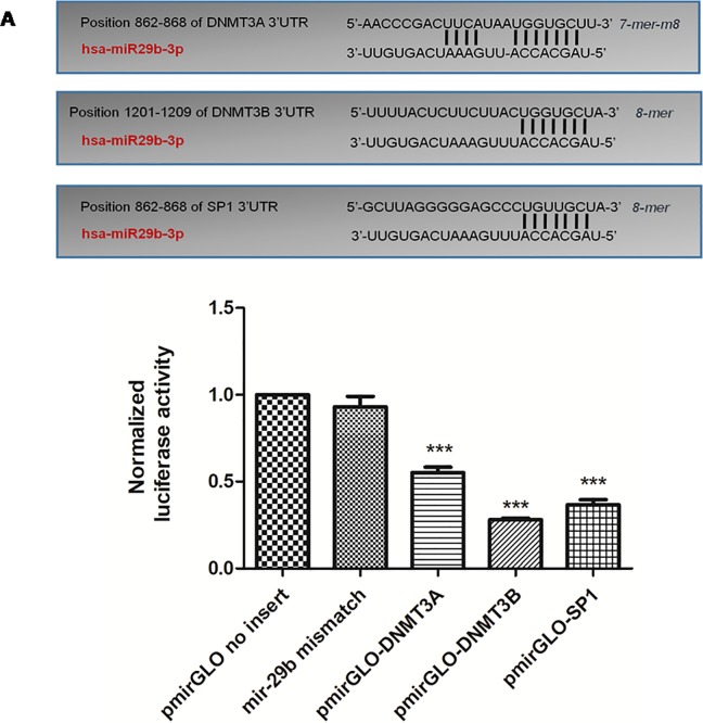
Fig 10. Luciferase activity measurement: The 3’-UTRs of DNMT3A, DNMT3B and SP1 are targets of miR-29b.
This figure describes a schematic of binding sites of miR29b with 3’-UTRs of DNMT3A, DNMT3B and SP1. Below is the graphical representation of Dual Luciferase Assay illustrated normalised Luciferase activity of pmirGLO vector (without 3’-UTR insert) and cells transfected with miR29b mismatch sequence (scrambled oligonucleotide). Further, pmirGLO vector with 3 different inserts of DNMT3A, DNMT3B and SP1 were allowed to interact with miR29b mimics independently and their Luciferase activity was examined as shown in this bar graph indicating significant difference in comparison with pmirGLO vector (without insert) and cells transfected with miR29b mismatch sequence (***p< 0.001).

