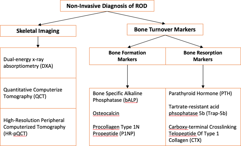
Figure 2. Non-Invasive Diagnosis of ROD.
Non-invasive skeletal imaging can be used to assess the presence of skeletal abnor-malities and to classify fracture risk. Measurement of bone mineral density can be done by dual energy X-ray absorptiometry (DXA). Microarchitecture and mineral density of trabecular and cortical bone can be assessed by quantitative computerized tomography (QCT) and by high-resolution peripheral computerized tomography (HR-pQCT).
Alternatively, assessment of turnover type can also be based on bone turnover markers. Bone formation markers, which are markers of osteoblast function, include bone specific alkaline phosphatase (BALP), osteocalcin, and procollagen type-1 N-terminal propeptide (P1NP). Bone resorption markers, which are markers of osteoclast number and function, include tartrate-resistant acid phosphatase 5b (Trap-5b) and C-terminal telopeptides of type I collagen (CTX).

