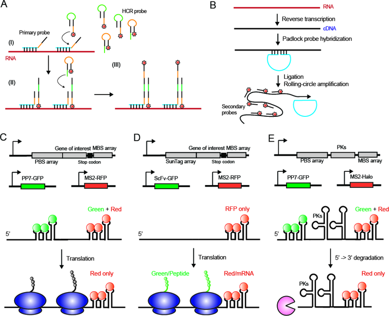
Figure 3: Emerging mRNA imaging methods in eukaryotic systems.
(A) In the in situ hybridization chain reaction (HCR), binding of the primary probe initiates the alternating binding of two HCR probes thereby amplifying the signal. (B) In the in situ polymerase chain reaction (PCR), a cDNA is first generated from the RNA of interest. Padlock probes are hybridized to the cDNA and ligated to be circular DNAs. Fluorophore labeled secondary probes are then hybridized to the products generated from rolling circle amplification of these circular DNA templates. (C) Schematic representation of the TRICK reporter construct. (D) Schematic representation of the SunTag construct. (E) Schematic representation of the TREAT reporter construct.

