
Figure 1.
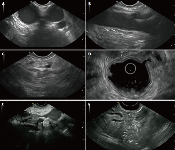
The endosonographic image of six characteristic views produced by a curvilinear array echoendoscope (Pentax EG3870UTK, Tokyo, Japan) and an ultrasound processor (Hitachi HI VISION Ascendus, Tokyo, Japan). Images by the authors. A: The aortopulmonary window (esophageal view); B: The abdominal aorta with the exit of the celiac trunc (gastric view); C: The left adrenal (gastric view); D: The stomach wall and its five layers (radial echoendoscope). Thickened wall (MALT-lymphoma) in the upper right part of the image; E: The pancreatic body including the splenic vein below (gastric view); F: The pancreatic head with the common bile duct and the pancreatic duct (duodenal view).