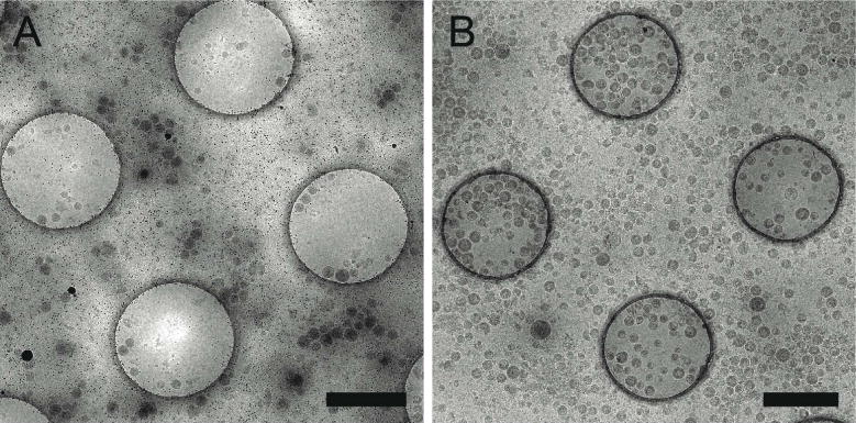
Figure 2. Affinity grid designed to selectively capture virus-like particles (VLPs).

Cryo-EM images of HIV CD84 VLPs applied to an untreated grid (A) and a 20% Ni-NTA cryo-affinity grid with His-tagged Protein A and anti-Env polyclonal antibody (B). Use of the affinity capture method leads to increased VLP concentration and improved particle distribution on the grid. See Kiss et al. (Kiss, et al., 2014) for experimental detail. Scale bars, 1 μm.
