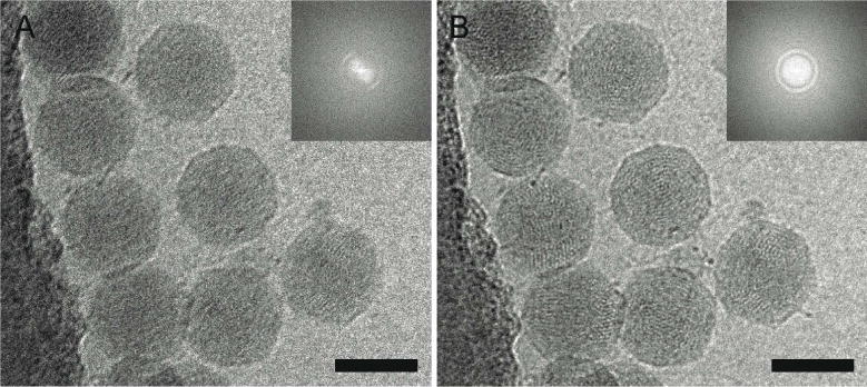
Figure 3. Motion correction of data recorded on a Direct Electron DE-20 direct electron detection device significantly improves image quality.

2D projection cryo-EM image of coliphage BA14 particles before (A) and after (B) motion correction using Direct Electron, LP scripts and the corresponding power spectra (insets). The image was recorded at a frame rate of 12 frames per second with an exposure time of 5 seconds. Scale bars, 50 nm.
