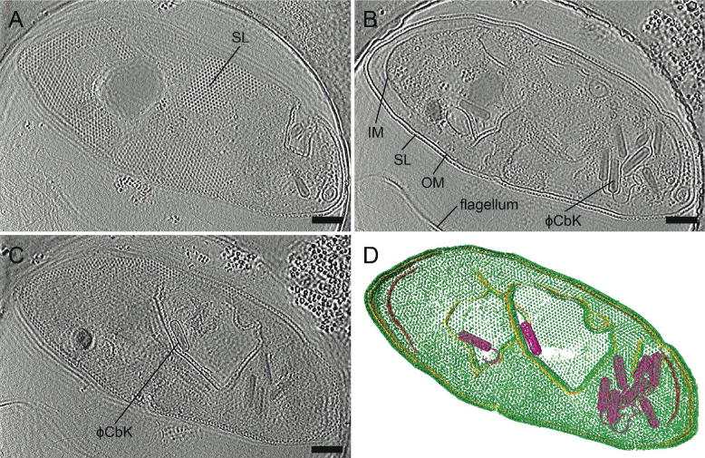
Figure 4. Zernike phase plate imaging of a phage-lysed bacterial cell provides contrast, revealing internal features.

Cryo-ET slices of ϕCbK phage-lysed Caulobacter crescentus cell using ZPC at zero defocus. (A) A top slice of the tomogram illustrating the hexagonal surface layer (SL), (B) a central slice revealing newly assembled phages within the lysing cell, and (C) a central slice showing an assembled phage capsid in the process of genome packaging. Fringing artifacts are evident, particularly at the edge of the cell. (D) Corresponding 3D segmentation showing surface layer, SL, green; outer membrane, OM, gold; inner membrane, IM, red; and ϕCbK, magenta. Scale bars, 200 nm.
