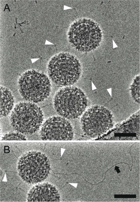
Figure 5. Hole-free phase plate (HFPP) imaging provides enhanced contrast without strong fringing artifacts.

Cryo-EM images of reovirus T1L particles using HFPP slightly underfocus. (A and B) reovirus T1L particles displaying attachment fibers as indicated by white arrowheads. The black arrow points to a released viral genome in (B). Scale bars, 50 nm.
