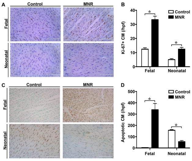
Figure 4. Maternal nutrient restriction increases the cardiomyocyte proliferative index in fetal and neonatal hearts.
A. Photomicrographs of ventricular myocardium from control and MNR-FGR fetuses and neonates stained with anti-Ki-67 antibody (brown) and counterstained with hematoxylin (blue). Scale bar equals 100μm. B. Quantification of Ki-67 positive cardiomyocytes (%) per 40X hpf in fetal and neonatal control (white bars) and MNR-FGR (black bars) hearts. Data represent mean number ± S.D. (n=6–8 per group, *p<0.001). C. Photomicrographs of ventricular myocardium from control and MNR-FGR fetuses and neonates stained with anti-TUNEL antibody (brown) and counterstained with hematoxylin (blue). Scale bar equals 100μm. B. Quantification of TUNEL positive cardiomyocytes (%) per 40X hpf in fetal and neonatal control (white bars) and MNR-FGR (black bars) hearts. Data represent mean number ± S.D. (n=4 per group, 1 animal per litter, *p<0.001). Analysis by 2-way ANOVA.

