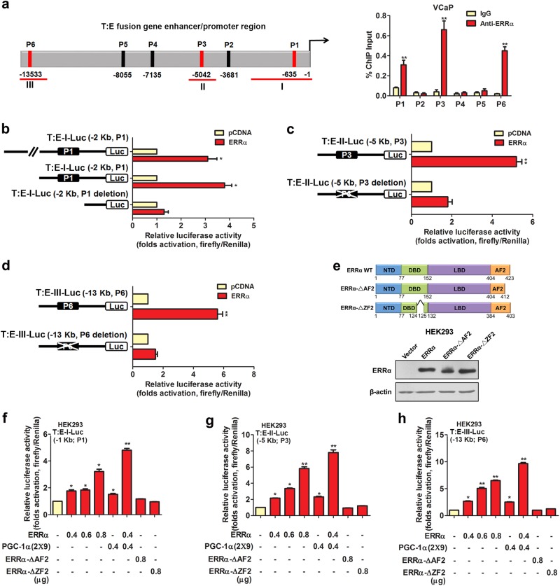
Fig. 5.
ERRα can directly transactivate the T:E fusion gene in AR-positive prostate cancer cells. a ChIP assay of T:E fusion gene performed in AR-positive VCaP cells. Left panel: schematic diagram shows the predicted ERRα-binding sites (P1–P6) located in the promoter and enhancer regions of T:E fusion gene. Right panel: results validated that three sites, namely P1, P3, and P6, located respectively at −640 bp, −5 kb and −13.5 kb upstream of the T:E fusion gene transcription start site, were enriched of ERRα. b–d Luciferase reporter assays of T:E fusion gene regulatory regions performed in ERRα-transfected HEK293 cells. All three T:E-I/II/III-Luc reporter constructs, driven by different lengths of T:E promoter/enhancer fragments, could be significantly transactivated by ERRα. Deletion of ERRα binding motifs in the T:E-Luc reporters abolished or significantly reduced the ERRα-induced transactivation. e Upper panel: Schematic diagram shows the domain structures of the wild-type ERRα protein and its truncated mutants. Lower panel: Immunoblot detection of transfected ERRα and its truncated mutants in HEK293 cells. (f–h) Luciferase reporter assays of T:E-I/II/III-Luc constructs performed in HEK293 cells. All three reporter constructs could be dose-dependently transactivated by ERRα, with further potentiation by co-transfection with PGC-1α (2 × 9), but not by ERRα-ΔAF2 and ERRα-ΔZF2 truncated mutant constructs. *P < 0.05; **P < 0.01 versus empty vector pcDNA3.1; two-tailed Student’s t test. All data are presented as mean ± SEM obtained from three independent experiments

