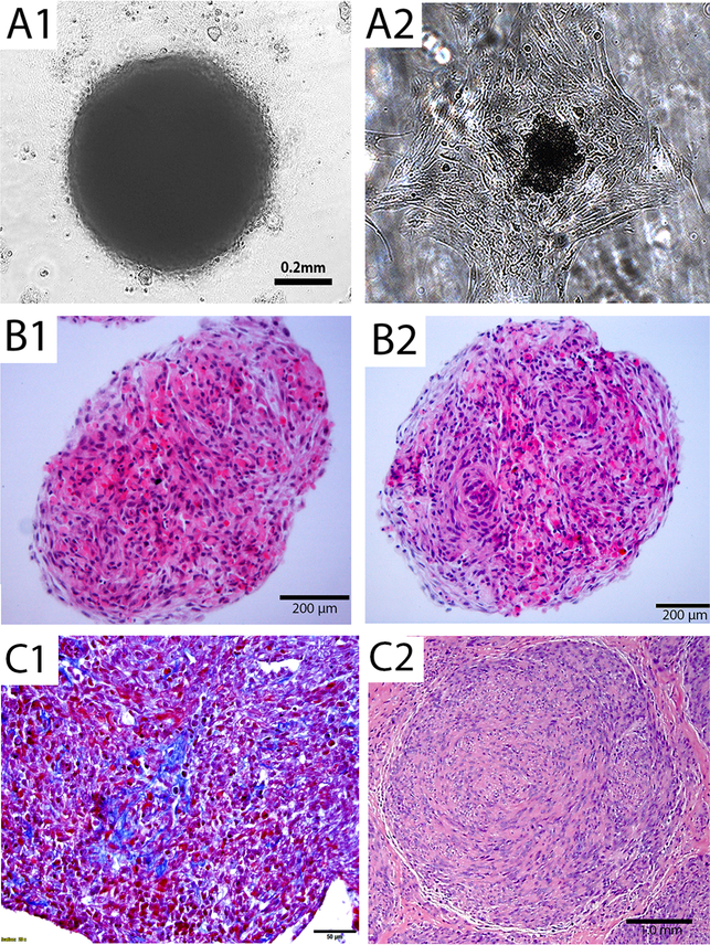
Figure 1. Three dimensional spheroid culture for ULM cells.
(A) 3D spheroid nodule formation in 3D suspension culture illustrated in an invert light microscope (A1) and 3D spheroid nodule can reattach to form monolayer after transferring to regular culture plate (A2). (B) Section of 3D spheroid nodules from myometrial (B1) and ULM (B2) on hematoxylin and eosin stained slides. (C) Trichrome stain shows thin and delicate collagen (C1). A small primary ULM (C2) from uterus was presented as a comparison. Black bars indicated amplification.

