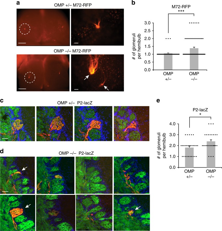
Fig. 5.
Duplicate glomeruli in the OMP–/– mice. a (Left) Whole mount of the OB dorsal surface in a M72-RFP OMP+/– (Top) and OMP–/– (Bottom) mouse with a the M72 glomerulus fluorescently labeled. Scale bar, 500 μm. (Right) Zoom-in of circled region. Note the duplicated glomeruli in the OMP–/– mouse (arrows). Scale bar, 100 μm. b Quantification of duplicate glomeruli in M72-RFP mice for OMP+/– and OMP–/– mice. Shown are 1.05 ± 0.03 glomeruli per hemifield (mean ± SEM, n = 56 hemibulbs) in OMP+/– mice and 1.36 ± 0.07 glomeruli per hemifield (mean ± SEM, n = 76 hemibulbs) in OMP–/– mice. c Serial coronal sections through individual glomeruli in one hemibulb of a P2-lacZ OMP+/– mouse. P2-positive OSN axons are labeled with β-Gal (red), OSN axons with SpH (green), and nuclei of juxtaglomerular cells surrounding glomeruli by DAPI (blue). Scale bar, 50 μm. d Serial coronal sections of a multiple glomeruli in one hemibulb of a P2-lacZ OMP–/– mouse. Arrows indicate distinct glomeruli and fluorescent labels as in (c). Scale bar, 50 μm. e Quantification of duplicate glomeruli in P2-lacZ mice for OMP+/– and OMP–/– mice. Shown are 1.82 ± 0.14 glomeruli per hemifield (mean ± SEM, n = 28 hemibulbs) in OMP+/– mice and 2.39 ± 0.16 glomeruli per hemifield (mean ± SEM, n = 28 hemibulbs) in OMP–/– mice. *P < 0.05, ***p < 0.001, Wilcoxon rank-sum test

