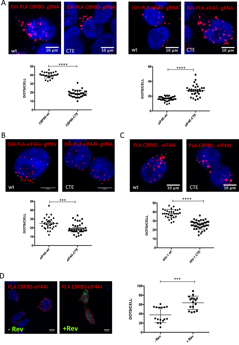
Figure 5.
The Rev/RRE axis favors the recruitment of CBP80 and eIF4AI to the HIV-1 unspliced mRNA. (A) HeLa cells were transfected with 1 μg pNL4.3-wt or 1 μg pNL4.3-CTE together with 1 μg of pCMV-myc-CBP80 or pCMV-myc-eIF4E. At 24 hpt, the interaction between the unspliced mRNA and CBP80 or eIF4E was analyzed by ISH-PLA. Scale bar 10 mm (upper panels). Dots per cell quantifications for total unspliced mRNA-CBP80 or unspliced mRNA-eIF4E interactions are presented in the lower panels. Dots quantifications were performed with ImageJ (****P < 0.0001, Mann–Whitney test). (B) HeLa cells were transfected with 1 μg pNL4.3-wt or 1 μg pNL4.3-CTE together with 1 μg of pCIneo-HA-eIF4AI. At 24 hpt, the interaction between unspliced mRNA and eIF4AI was analyzed by ISH-PLA. Scale bar 10 mm (upper panel). Dots per cell quantifications for total unspliced mRNA-eIF4AI interactions are presented in the lower panel. Dots quantifications were performed with ImageJ (***P < 0.001 and NS; non-significant, Mann–Whitney test). (C) HeLa cells were transfected with 1 μg pNL4.3-wt or 1 μg pNL4.3-CTE together with 1 μg pCMV-myc-CBP80 and 1 μg pCIneo-HA-eIF4AI. At 24 hpt, the interaction between HA-eIF4AI and myc-CBP80 was analyzed by PLA (upper panels). Scale bar 10 μm. Dots per cell quantification of eIF4AI and CBP80 interactions were performed with ImageJ (****P < 0.0001, Mann–Whitney test). (D) HeLa cells were transfected with 1 μg pCMV-myc-CBP80 and 1 μg pCIneo-HA-eIF4AI together 1 μg pEGFP-Rev (pEGFP was used as a control). At 24 hpt, the interaction between HA-eIF4AI and myc-CBP80 was analyzed by PLA (left panels). Scale bar 10 μm. Dots per cell quantification of eIF4AI and CBP80 interactions were performed with ImageJ (***P < 0.001, Mann–Whitney test).

