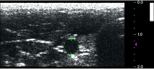
Figure 1. Measurement of vessel luminal area.

Measurements of the arterial lumen area were obtained using cine axial images with focus zone set at the level of the vessel and depth of 2 cm using the built‐in software. The arteries were differentiated from the adjacent veins by either the presence of visible pulsatile movement or arterial Doppler waveform evaluations. [Color figure can be viewed at http://wileyonlinelibrary.com]
