
Figure 1. The processes determining placental amino acid transfer operate across a range of scales.
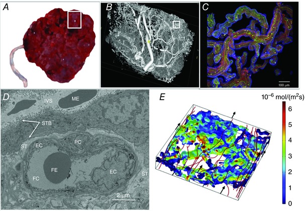
Amino acids within maternal blood mix effectively between the placental villi before being transported across the apical and basal membranes of the syncytiotrophoblast, diffusing through the stroma and then through the junctions between endothelial cells. A, a whole placenta approximately 20 cm across. The white box indicates one placental lobule (∼3 cm across). B, micro‐computed tomography image of the region shown in the white box in A, illustrating the extent and complexity of the fetal vasculature. The white box in B indicates a region of placental villi. C, the region shown by the white box in B shown as a projection of a whole mount confocal image stack, with the trophoblast stained blue, connective tissue red and endothelium green. D, an electron microscopy image of a cross section of a terminal villi. E, computational 3D simulation results modelling streamlines of maternal blood flow (m s−1) through the intervillous space surrounding placental villi, with streamlines in red depicting the main flow routes (flow direction indicated by black arrows). Colour on the villous surface represents uptake of substrate in that area with red indicating highest solute flux through the villous barrier (mol m−2 s−1); predicted using the computational model. EC, endothelial cell; FC, fetal capillary; FE, fetal erythrocyte; IVS, intervillous space; ME, maternal erythrocyte; PC, pericyte; ST, villous stroma; STB, syncytiotrophoblast (the microvillous membrane and basal plasma membranes of the syncytiotrophoblast are indicted by the white arrows).