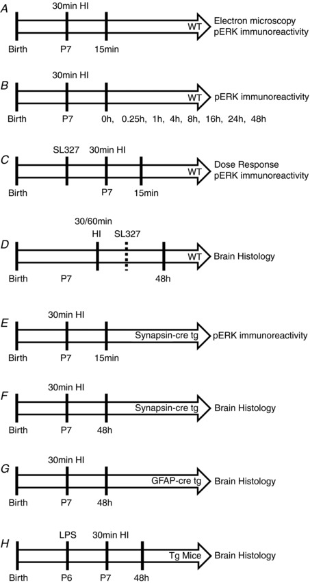
Figure 1. Schedule of experimental procedures.

A, WT (C57/Bl6) mice underwent 30 min hypoxic–ischaemic (HI) insult and were then killed at 15 min post‐hypoxia for pERK immunoreactivity evaluation. B, pERK immunoreactivity was assessed at multiple time points up to 48 h post‐insult to P7 WT mice. C, a dose response of SL327, controlled to vehicle alone, was administered 20 min prior to 30 min HI and pERK immunoreactivity was assessed at 15 min post‐insult. D, WT mice were subject to either 30 min or 60 min HI, with 133 μg/g SL327 or EtOH (vehicle) administered either 20 min prior to or 60 min post‐insult. Brain histology was assessed at 48 h. E, inhibition of neuronal pERK immunoreactivity was confirmed at 15 min post‐HI in synapsin‐cre driven ERK tg mutant mice compared to littermate WT controls. F, brain histology was assessed at 48 h after 30 min HI in synapsin‐cre driven ERK tg mutant mice and littermate WT controls. G, brain histology was assessed at 48 h after 30 min HI in GFAP‐cre driven ERK tg mutant mice and littermate WT controls. H, saline or LPS was injected at 12 h prior to 30 min HI in both synapsin‐cre and GFAP‐cre driven ERK tg mutant mice and littermate WT controls. Brain histology was assessed at 48 h.
