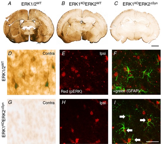
Figure 6. Effects of global ERK1 and neuronal ERK2 deletion on pERK immunoreactivity at 15 min post‐30 min HI insult.

A–C, control (ERK1/2WT) animal (A), global deletion of ERK1 and ERK2WT (ERK1KO) (B), global ERK1 deletion and homozygous neuronal ERK2 deletion (ERK1KOERK2ΔSyn) (C). C, pERK immunoreactivity is almost completely reduced following deletion of both copies of ERK1 and ERK2. D–I, quantification and distribution of pERK immunoreactivity at high magnification. D, pERK staining in the contralateral pyriform cortex of ERK1/2WT with strong neuronal reactivity and prominent dendritic staining which disappears in the presence of global ERK1 deletion and homozygous neuronal ERK2 mutation ERK1KOERK2ΔSyn. E–I, residual immunoreactivity on the ipsilateral side. E and H, pERK alone. F and I, immunofluorescence double labelling with GFAP demonstrating co‐localisation of pERK in astrocytes, particularly pronounced in ERK1WTERK2ΔSyn (white arrows). Scale bar = 25 µm [Color figure can be viewed at http://wileyonlinelibrary.com]
