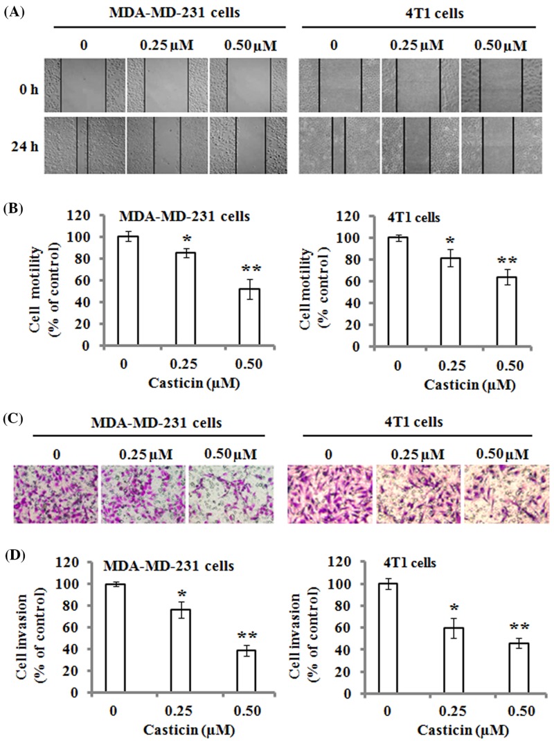
Figure 2. Effects of casticin on cell migration and invasion.
The monolayers of MDA-MB-231 and 4T1 cells were respectively scratched with a pipette tip, and incubated with 0, 0.25, and 0.50 µM of casticin for 24 h. (A) Representative images of wound healing. Original magnification was ×100. (B) The number of cells migrated to the denuded zone was quantitated, and normalized to that of the control. (C) Cell invasion was analyzed with a Matrigel-coated Boyden chamber. MDA-MB-231 and 4T1 cells were respectively treated with 0, 0.25, and 0.50 µM of casticin for 24 h, and then analyzed as described in the ‘Materials and methods’ section. Representative photomicrographs of the membrane-associated cells were assayed by H&E staining. Original magnification was ×400. (D) Cell invasion ability was quantitated. Data represent the mean ± S.D. of three independent experiments. *P<0.05 and **P<0.01 compared with the control.

