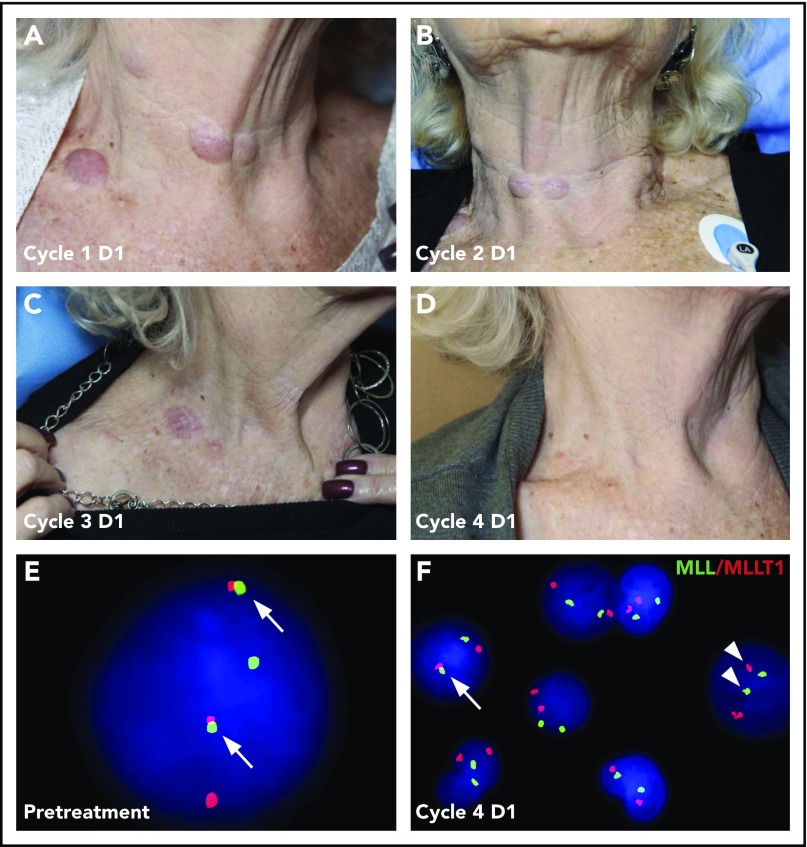
Figure 3.
Resolution of leukemia cutis and cytogenetic changes following pinometostat treatment. Cutaneous leukemia cutis in an 81-year-old patient presenting with MLL-r CMML that was treated with 54 mg/m2 per day of pinometostat by 21-day CIV infusion. Leukemia cutis neck lesions that were apparent on day 1 (D1) at the start of treatment (A) progressively resolved over the course of subsequent treatment cycles (B-D). (E) Translocation-positive cell in a peripheral blood sample detected by FISH from the same patient showing the t(11:19) MLL-r product colocalizing with the MLLT1 fusion partner (arrows). (F) The number of translocation-positive cells (arrow) decreased from 90% pretreatment to 0.2% at the start of the fourth pinometostat treatment cycle, with most cells demonstrating normal segregation of MLL and MLLT1 signals (arrowheads).

